Can U Have a Ct Scan After a Hip Replacement
Computed Tomography [edit | edit source]
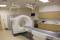
Computed tomography (CT) is an imaging procedure that uses special x-ray equipment to create detailed pictures, or scans, of areas inside the body. Information technology is as well chosen computerized tomography and computerized axial tomography (CAT). The termtomography comes from the Greek wordstomos (a cut, a slice, or a section) andgraphein (to write or tape).[1] Computed tomography (CT) is noninvasive and produces cantankerous-sectional images of the trunk. Each cross-exclusive image represents a "slice" of the person being imaged, similar the slices in a loaf of staff of life. These cross-sectional images are used for a variety of diagnostic and therapeutic purposes.[ii]
CT scans tin exist performed on every region of the body for a diversity of reasons (e.g., diagnostic, treatment planning, interventional, or screening). Most CT scans are performed as outpatient procedures. Computed tomography was originally known as the "EMI scan" as information technology was developed at a research branch of EMI, a visitor best known today for its music and recording business organisation. It was later known as computed axial tomography (Cat or CT scan) and body section röntgenography.
The cantankerous-sectional images generated during a CT scan tin be reformatted in multiple planes, and can even generate three-dimensional images which can be viewed on a computer monitor, printed on film or transferred to electronic media.[3]
Although most common in medicine, CT is also used in other fields, such as nondestructive materials testing. Some other example is the DigiMorph projection at the Academy of Texas at Austin which uses a CT scanner to study biological and paleontological specimens.
Purpose [edit | edit source]
CT-scans provide detailed cantankerous-exclusive images of various internal structures for example internal organs, blood vessels, bones, soft tissue etc, and can be used for:
- Diagnostic purposes-
- Guidance for specific treatment or further tests- surgeries, biopsies and radiations therapy
- Detection and monitoring of conditions- Cancer, heart disease, lung nodules, liver masses
Indications for CT Scans:
- Traumatic injuries
- Degenerative conditions, such as stenosis and osteoarthritis when an MRI is contraindicated
- Postal service-operative conditions
- Neoplastic conditions
- Infectious processes
- Image guidance during injections, biopsy's and aspirations
- Abnormalities of bony alignment, such equally scoliosis
- Processes involving the spinal cord when MRI is contraindicated[4]
Technique [edit | edit source]
Most modernistic CT machines take continuous pictures in a helical (or spiral) fashion rather than taking a serial of pictures of private slices of the body, as the original CT machines did. Helical CT has several advantages over older CT techniques: information technology is faster, produces better 3-D pictures of areas inside the body, and may observe small-scale abnormalities better. The newest CT scanners, called multislice CT or multidetector CT scanners, allow more slices to be imaged in a shorter catamenia of time.[1]
Digital geometry processing is used to generate a three-dimensional paradigm of the within of an object from a large series of two-dimensional Ten-ray images taken around a single axis of rotation. CT produces a book of data which can be manipulated, through a process known every bit "windowing", in order to demonstrate diverse actual structures based on their power to block the X-ray/Röntgen axle. Although historically the images generated were in the axial or transverse plane, orthogonal to the long axis of the torso, modernistic scanners let this volume of data to be reformatted in various planes or even as volumetric (3D) representations of structures.
Sometimes, CT involves the use of a dissimilarity (imaging) agent, or "dye." The dye may be given past mouth, injected into a vein, given by enema, or given in all three ways earlier the procedure. The contrast dye highlights specific areas inside the body, resulting in clearer pictures. Iodine and barium are ii dyes commonly used in CT[1]
Procedure[2] [edit | edit source]
- A motorized table moves the patient through a circular opening in the CT imaging system.
- While the patient is inside the opening, an X-ray source and a detector assembly within the organization rotate around the patient. A unmarried rotation typically takes a second or less. During rotation the X-ray source produces a narrow, fan-shaped axle of X-rays that passes through a department of the patient'due south body.
- Detectors in rows opposite the Ten-ray source register the X-rays that pass through the patient's body as a snapshot in the procedure of creating an image. Many different "snapshots" (at many angles through the patient) are nerveless during one complete rotation.
- For each rotation of the X-ray source and detector assembly, the image data are sent to a reckoner to reconstruct all of the individual "snapshots" into 1 or multiple cross-sectional images (slices) of the internal organs and tissues.
Resources [edit | edit source]
[5]
References [edit | edit source]
- ↑ one.0 1.one 1.two National Cancer Found. Computed Tomography (CT) Scans and Cancer [Internet]. 2022 [cited xxx/04/2019]. Bachelor from: https://www.cancer.gov/near-cancer/diagnosis-staging/ct-scans-fact-sheet.
- ↑ 2.0 2.ane U.Southward Food and Drug Administration. Computed Tomography (CT) [Internet]. 03/06/2018 [cited xxx/04/2019]. Bachelor from: https://world wide web.fda.gov/radiation-emitting-products/medical-x-ray-imaging/computed-tomography-ct
- ↑ Radiologyinfo.org. Computed Tomography [Internet]. 2022 [cited xxx/04/2019]. Available from: https://www.radiologyinfo.org/en/submenu.cfm?pg=ctscan
- ↑ College, A. ACR – ASNR – ASSR – SPR Exercise GUIDELINE FOR THE PERFORMANCE OF COMPUTED TOMOGRAPHY ( CT ) OF THE SPINE. Diagnostic Imaging, one-7. 2011
- ↑ The National Institute of Biomedical Imaging and Bioengineering. How Does a CT Scan Piece of work? [Internet]. 2022 March 7. [cited 2022 April thirty]. Bachelor from: https://world wide web.youtube.com/sentinel?v=l9swbAtRRbg
Source: https://www.physio-pedia.com/CT_Scans
0 Response to "Can U Have a Ct Scan After a Hip Replacement"
Post a Comment